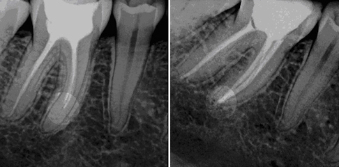
The left picture illustrates a complaints causing, elsewhere root canal filled tooth contacting us patient’s before treatment X-rays. The right picture is about the same tooth, it is made after the removal of the old root canal filling and the new root canal filling preparation.
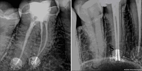
The preparation of a precise root canal filling wishes unfortunately time, practice and proper instrumentation, but the good news is that it is painless. The mechanical root canal reamers, the distance from the root apex continuously measuring instruments, in the chair during the treatment can be made digital high resolution radiographs which greatly help the doctor’s work. Particularly thin and curved roots’ filling is “art”, such as on the left picture. On the right picture an own (s) and away (i) made root canal filling can be seen.
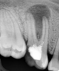
The picture below shows an inadequate root canal filling causing cyst’s at the root apex.